
• Home
  • NAMs & SVC
  • サービス-NeXST SVC
  • Cardiac contractility

Cardiac contractility

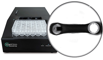
Mantarray

Mantarray
  • Real-time 3D forces
  • Label-free, magnetic sensor based
  • Electric pacing

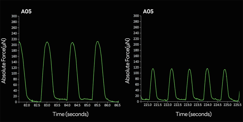
Mantarray Baseline vs 48-hours

  • Doxorubicin 1 μM, Chronic Treatment

3D Engineered Heart Tissue (EHT)

- Contraction force of cardiomyocytes is measured using 3D engineered heart tissue.
- Aligned cardiomyocyte arrangement
- Enhanced cardiomyocyte maturation
- Stronger contractile force
- More accurate drug responsiveness compared to 2D cardiomyocytes

Applications

- Screening of drug-induced changes in cardiac contractility (from hit compounds to preclinical stage)
- Evaluation of efficacy and toxicity of candidate drugs

Flexcyte

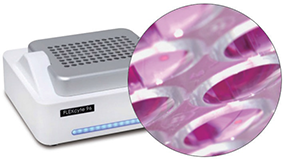
FLEXcyte 96
  • Physical contractility
  • Flexible membrane bottom
  • Optical pacing
  • High-throughput

FLEXcyte96 Compound Treatments
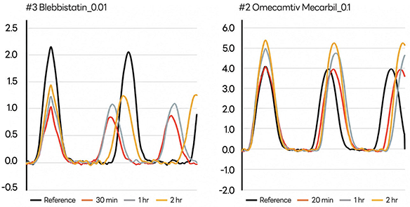
  • Blebbistatin 0.01 μM, Myosin II ATPase Inhibitor
  • Omecamtiv Mecarbil 0.1 μM, Myosin Activators

“Flexcyte Contractility Service – Evaluating Drug Responses in a Heart-like Environment“

NeXST’s Flexcyte-based Contractility Service enables quantitative measurement of mechanical contraction force in hiPSC-derived cardiomyocytes under physiologically relevant conditions that closely mimic the human heart.
The FLEXcyte 96 platform cultures cells as monolayers on ultra-thin, flexible polydimethysiloxane (PDMS) membranes, allowing the synchronized beating of cardiomyocytes to be directly recorded as true contractile force. This unique approach captures even subtle drug-induced changes that conventional electrophysiology may miss.

·Key Advantages
1. High-throughput capability (96-well format) – parallel recordings from dozens of compounds
2. Clinically relevant insights – data generated in a native-like mechanical environment
3. Versatile applications – cardiotoxicity and safety screening, efficacy profiling, and patient-derived cell research

With Flexcyte, NeXST goes beyond electrical readouts to deliver comprehensive and reliable cardiac safety evaluation based on true mechanical contractility.

“Uncovering Drug-Induced Cardiac Contractility Changes with Flexcyte”

With the Flexcyte platform, it is possible to quantitatively analyze drug-induced changes in cardiomyocyte contractility.
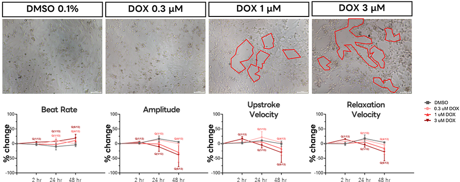
For example, treatment with the anticancer agent Doxorubicin allows real-time monitoring of critical parameters such as beat rate, amplitude, upstroke velocity, and relaxation velocity, along with simultaneous observation of morphological alterations.

Acute: Result of Verapamil treatment

Contractility changes of Cardiosight®-S in response to the Ca2+ channel blocker verapamil, as measured using the FLEXcyte96 system (Acute).

(A) Representative traces after treatment with 0.1% DMSO (NC) and Verapamil. The NC 0.1% DMSO shows no significant changes for up to 2 hours, while verapamil demonstrates a dose-dependent decrease in contractility.

(B) Bar graph showing changes according to concentration and time. Overall parameters show a dose-dependent decrease, but a time-dependent recovery of cells is observed

Chronic: Result of Doxorubicin treatment

Contractility changes of Cardiosight®-S in response to doxorubicin, a death receptor-mediated apoptosis inducer, as measured using the FLEXcyte96 system (Chronic).

(A) Morphology of Cardiosight®-S on the FLX-96 plate after 48 hours of treatment with 3 μM Doxorubicin (DOX). The toxicity of DOX weakens cell-cell junctions, and a dose-dependent increase in hole size is observed.

(B) Time- and dose-dependent decreases in parameters are observed. Cell activity decreases due to cytotoxicity, with all parameters except beat rate showing a reduction. Additionally, there is a dose-dependent increase in the number of wells exhibiting quiescence.

Muscle motion

“Simple Video-based iPSC-CM Contractility Analysis – Muscle motion program”

The Muscle Motion program is an innovative tool that enables precise analysis of iPSC-derived cardiomyocyte contraction using only simple cell culture videos. Without the need for specialized equipment or complex setups, videos recorded from either standard culture plates or specialized plates can be directly analyzed, making it a cost-effective and highly accessible solution.

·Key Advantages
1. Low-cost, high-efficiency cardiomyocyte contractility assessment
2. Compatible with both standard and specialized plates
3. Supports long-term culture-based contraction analysis

The Muscle Motion Program enhances research efficiency while expanding the accessibility and applicability of iPSC-CM–based cardiac contractility evaluation.

Cell video analysis with Image J software

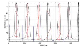
Contraction Analysis

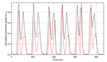
Speed of Contraction Analysis

Analysis parameter: Beat rate, Contraction amplitude, Rise rate, and Decay rate

Beyond conventional boundaries,
we deliver a bold new paradigm.