Organoid | 3D Heart Organoids
Human induced Pluripotent Stem Cells(hiPSCs) derived Cardiac & Heart Organoids
Differentiation of Cardiac & Heart Organoid
Overall schematic diagram of differentiation from hiPSC into COs and HOs.
Specifications of Cardiac & Heart Organoid
| |
COs |
HOs |
| Organoid Diameter |
200 ~ 400 μm |
600 ~ 800 μm |
Cardiomyocytes distribution
(cTnT positive)
|
± 95% |
± 50 ~ 60% |
Sarcomere length
(α-actinin positive)
|
± 15 μm |
Cell type composition
(scRNA-seq check)
|
Cardiomyocytes |
Cardiomyocytes
Fibroblasts
Endothelial cells
|
| Beating efficacy |
Beating efficacy |
Spike amplitude
(measured by MEA)
|
± 0.72 mV |
± 0.38 mV |
| BPM |
60 ~ 80 |
50 ~ 60 |
Cardiac Organoid Morphology & Drug
Response Evaluation
Using an optimized proprietary protocol, we have created high-quality human cardiomyocytes differentiated from iPSCs in the form of cardiac organoids.
These cardiac organoids exhibit dose-dependent responses to standard drugs.
Heart Organoid Structural Characteristics
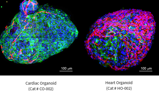
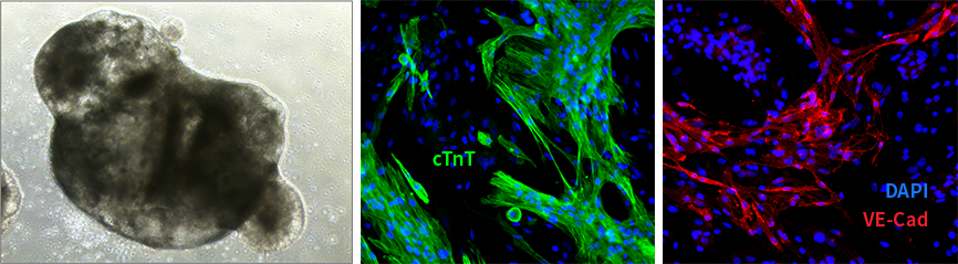
The heart organoid displays a three-dimensional, spherical structure that resembles the architecture of the developing heart.
The ICC for cardiac troponin T (cTnT) shows the expression of this key cardiac muscle marker within the heart organoid.
The ICC for vascular endothelial cadherin (VE-Cad) demonstrates the presence of endothelial cells within the heart organoid, highlighting the structural and cellular complexity of cardiac organoids.
Heart Organoid disease modeling
Schematic diagram the implementation of heart disease occurring in vivo to in vitro organoids
Structural Defects in Disease-Cultured Organoids
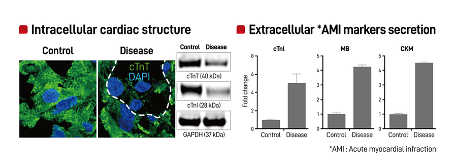
The data presented shows the release of cardiac-specific markers, such as cTnT and cTnI, which are indicative of acute
myocardial infarction (AMI) in the organoids.
The three markers, cTnI (Cardiac troponin I), MB (Myoglobin), and CKM (Creatine kinase), are known to
be present at high concentrations in the blood when a myocardial infarction occurs in a clinical setting.
Therefore, the observation of these myocardial infarction-specific markers being released in the organoids with induced myocardial infarction suggests
that the organoids are exhibiting similar characteristics to what is observed in the clinical setting.
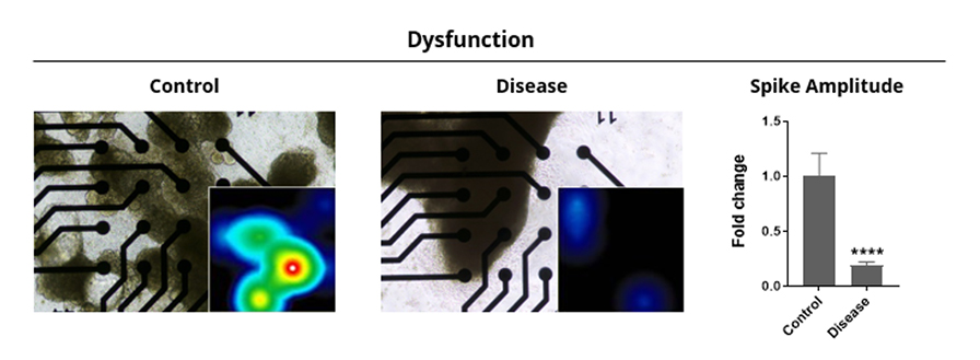
The electrophysiological measurements of organoids cultured in a disease environment show a decrease in electrical signals. One of the key functions of
the heart is the maintenance of beating through electrical signals.
The results demonstrate that the occurrence of a disease leads to a decrease in electrical signals, which in turn affects the heart's beating, as observed in cardiac organoids.





 CONTACT